
Page 39 - Biology - XII
P. 39
Identifi cation
This is the T.S. of mammalian testis (mouse).
Comments
1. Many oval-shaped structures are seen which are called seminiferous tubules.
2. Seminiferous tubules are lined by germinal epithelium which produce spermatogonia.
3. Various stages of spermatocytes (primary, secondary), spermatids, and spermatozoan are seen inside
the tubules.
4. Large cells called sertoli cells are there. These cells are seen in between germinal epithelium.
5. These sections of seminiferous tubules are surrounded by connective tissues, blood vessels, and nerve fi bres.
6. Also present are interstitial cells. Leydig cells outside the seminiferous tubules secrete male sex
hormone—testosterone.
SPOT - 2
T.S. of Mammalian Ovary (Mouse)
Identifi cation
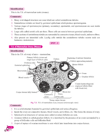
This is the T.S. of ovary of mice—mammalian.
Primary follicle (ovum and Beginning of antrium formation
single layer of follicle cells) Follicle Follicle approaching maturity
Oviferous tube Egg nest
Germinal epithelium Mature follicle
Mesovarium
Ovum
Blood vessels Graafi an follicle
Corpus haemorrhagicum
(ruptured follicle fi lled
with blood clot)
Corpus luteum fully formed Blood clot
Connective Fibrin
tissue Luteum cells
Young corpus luteum
Fig. 7.2: T.S. of mammalian ovary (mouse) (microscopic view)
Comments
1. It is a solid structure bounded by germinal epithelium and tunica albuginea.
2. Inside the ovary are connective tissues, blood vessels, nerve fi bres, etc. This forms the stroma of ovary.
3. Spherical/oval structures of various sizes called (ovarian) follicles are seen.
4. A mature follicle is called graafi an follicle. It is identifi ed by the presence of an ovum surrounded by a
group of follicular cells and follicular fl uid.
5. A spot of rupture of ovarian membrane is seen which later transforms into corpus luteum.
38