
Page 53 - Biology - XII
P. 53
EXPERIMENT - 9
Objective
To study the blastula stage of embryonic development in mammals with the help of permanent slides.
Principle/Theory
Embryo is the developing stage of the structure formed after zygote development. A single-celled zygote
gets transformed into a multicellular structure embryo. Diff erent stages of embryo development are: Morula
a solid cells structure followed by Blastula with a cavity called blastula and then gastrula that has three
germ layers which forms the organs in the developing embryo.
1. Embryonic development takes place by cleavage division that occurs in quick succession without any
synthetic phase, i.e. it is rapid process.
2. A zygote → blastomeres. The 16-celled stage of embryo is called morula followed by blastula.
(32-celled stage and then gastrula where three primary germinal layers are formed.)
Zygote Morula Blastula Gastrula
Materials Required
Permanent slides and compound microscope
Procedure
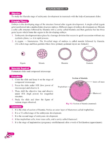
1. Clean the slide and keep it on the stage of Section of blastula
compound microscope. Inner cell mass
2. Focus the slide under 10X (low power of
microscope) and observe it. Trophoblast
3. Then, shift the objective lens and observe
under 45X (high power) for magnifi ed Blastocoel
structure.
4. Study the slide and draw the fi gure of
various stages observed.
Fig. 9.1: Section of blastula (mammal)
Comments
1. It is the slide of section of blastula. Notice an outer layer of blastomers called trophoblast.
2. It is a 32-celled stage of the embryonic development.
3. It is the second stage of embryonic development.
4. It has trophoblast cells, inner mass cells, and a cavity called blastocoel.
5. It is the stage of implantation (pregnancy establishment) after a week of fertilisation (approximate).
52